
High-quality imaging, close to home.
Cutting-edge technology in a comfortable, friendly environment. Every study is read by a U.S. board-certified radiologist who specializes in that body part, with results within 24 hours or STAT when needed.
Open MRI
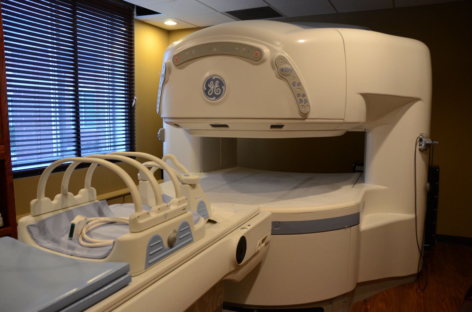
Our GE Signa HD Open MRI captures high-quality images while staying open on the sides, so you never feel closed in. Certified MRI technologists stay in constant communication with you throughout your scan.
MRI of soft tissue such as the heart, liver and other organs is often more likely to find and accurately characterize disease than other methods, which makes it invaluable for early diagnosis.
What is MRI?
Magnetic resonance imaging (MRI) is a noninvasive test that uses a powerful magnetic field, radio-frequency pulses and a computer to produce detailed pictures of organs, soft tissue, bone and other internal structures. MRI does not use ionizing radiation (X-rays).
How should I prepare?
- Wear loose, comfortable clothing without metal fasteners, or change into a gown.
- Unless told otherwise, eat and take your medications as usual.
- Tell us about allergies, kidney problems, pregnancy, or any implanted devices or metal in your body, such as a pacemaker, defibrillator, cochlear implant, aneurysm clips or shrapnel.
- Leave jewelry, watches, hearing aids and other metal items at home.
- If you are anxious or claustrophobic, you can ask your doctor about a mild sedative before your exam.
Please fill out the MRI patient forms before your visit.
What happens during the exam?
You lie on a padded table that moves into the open scanner. The machine makes loud knocking sounds while it takes pictures, so we offer earplugs and music. Some exams use a contrast injection through an IV. Most scans take 15 to 45 minutes, and the technologist can see, hear and speak with you the whole time.
How do I get my results?
A U.S. board-certified radiologist who specializes in the body part being studied reviews your images and sends a signed report to your doctor within 24 hours, or STAT when needed. Your doctor will share the results with you.
CT Scan
CT (sometimes called a CAT scan) is one of the best and fastest tools for examining the chest, abdomen and pelvis. It provides detailed, cross-sectional views of organs, bones, soft tissue and blood vessels, with more detail than a regular X-ray.
What is CT used for?
- Detecting many cancers and measuring their size and location
- Finding blood clots in the lungs and aneurysms
- Spine problems and injuries to small bones in the hands and feet
- Planning and checking the results of surgery and other treatments
- Measuring bone density to detect osteoporosis
How should I prepare?
- Wear comfortable, loose clothing. You may be given a gown.
- Leave jewelry, eyeglasses, hairpins and other metal at home.
- You may be asked not to eat or drink for a few hours beforehand if contrast will be used.
- Tell us about any allergy to contrast ("dye"), and about diabetes, asthma, heart, kidney or thyroid problems.
- Tell us if you are, or could be, pregnant.
- If you are over 60, diabetic, or have a history of kidney disease, bring BUN and creatinine (or GFR) lab results from the past 60 days.
- If you are allergic to iodine, your doctor may prescribe a 13-hour pre-medication prep.
Please fill out the CT patient forms before your visit.
What happens during the exam?
You lie on the table, which moves slowly through the opening of the scanner. You may be asked to hold your breath for a few seconds. The scan is painless and usually finished within 30 minutes. If contrast is used, you may feel warm briefly or notice a metallic taste for a minute or two.

Ultrasound
Ultrasound is safe and painless and makes pictures of the inside of the body using sound waves, with no radiation. Because the images are captured in real time, they show the structure and movement of organs and blood flowing through blood vessels.
What is a Doppler ultrasound?
A Doppler study is a special ultrasound technique that evaluates blood flow through the major arteries and veins of the abdomen, arms, legs and neck. You may hear pulse-like sounds during the exam.
How should I prepare?
Wear comfortable, loose clothing. Preparation depends on the exam: for some you may be asked not to eat or drink beforehand, and for others to drink several glasses of water and arrive with a full bladder. We'll tell you exactly what to do when you book. Please fill out the ultrasound patient forms before your visit.
What happens during the exam?
You lie on the exam table while the sonographer applies warm gel and moves a small handheld probe over the area. Most exams take 30 minutes to an hour, and you can go back to normal activities right away.

X-Ray
An X-ray is a quick, painless test that produces images of the structures inside your body, especially your bones. Dense materials like bone show up white, air in the lungs appears black, and fat and muscle appear in shades of gray.
Why is it done?
- Bones: fractures, infections, arthritis and bone density.
- Chest: pneumonia and other lung conditions, or an enlarged heart.
- Abdomen: digestive problems and swallowed objects.
How should I prepare?
Most X-rays need no preparation. Wear comfortable clothing; you may be asked to remove jewelry or change into a gown. Tell us if you are, or could be, pregnant. Please fill out the X-ray patient forms before your visit.
What can I expect?
You'll be positioned on the table or against the stand, and each image takes only seconds. You may be asked to hold still or hold your breath briefly. Most X-ray exams take just a few minutes.

MRI Arthrograms
An MRI arthrogram takes detailed pictures of a joint in two parts: first contrast is injected into the joint, then an MRI is performed. Together they show more detail than an MRI alone and help your doctor choose the best treatment. The exam takes about 30 minutes plus your MRI.
Why am I having an arthrogram?
Joints such as the knee, hip, shoulder and wrist are supported and cushioned by cartilage, tendons and ligaments. An arthrogram helps your doctor see small tears or other problems in these tissues and track them over time.
How should I prepare?
Please call at least 24 hours before your exam if you need to reschedule, or if any of these apply:
- Allergy to contrast (X-ray dye, iodine or MRI contrast)
- Diabetes
- Taking warfarin (Coumadin) or other blood thinners
- Being treated for an infection
- Claustrophobia
Bring a list of all your medicines. You will give written consent before the exam begins.
Please fill out the MRI arthrogram patient forms before your visit.

Post Discograms
A discogram helps your doctor find out whether a specific disc in your spine is causing your back pain. Contrast is injected into the soft center of the disc, then an X-ray or CT scan shows whether it has spread through cracks in the disc's outer layer.
How should I prepare?
You may need to stop blood-thinning medicines for a period of time beforehand; your doctor will tell you which medicines you can take. Do not eat or drink the morning of the test.
What will I experience?
You lie on your stomach while the doctor numbs the skin and guides a thin needle into the disc using live X-ray. The injection may briefly reproduce your usual back pain, and you'll be asked to rate it. Some soreness for a few hours afterward is normal; keep your back dry for 24 hours.
How do I get my results?
Your doctor reviews the images together with what you felt during the injection to pinpoint the source of your pain and plan your treatment.

Questions about your exam?
Our staff is happy to help you prepare.